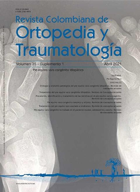
Prevención, identificación y tratamiento de las recidivas en el pie equino varo congénito. Revisión de conceptos actuales
DOI:
https://doi.org/10.1016/j.rccot.2021.01.002Palabras clave:
prevención, recidiva, recaída, talipes equinovaro, pie equino varo, pie bot, pie zambo, cirugía ortopédica, tenotomía, tendón de aquiles, férula, transferencia tendinosa, deformidades del pie, factores de riesgoResumen
Se entiende por recidiva la aparición progresiva de las deformidades del pie equino varo congénito y suele manifestarse por lo común durante los primeros cinco años de vida del niño. La principal causa de la recidiva es el uso inadecuado de la férula de abducción que tiene la función de estirar los tejidos blandos posteriores e internos de la pierna y el pie. Por esta razón, es fundamental una comunicación estrecha del médico con los padres o acudientes del niño para hacerles entender la importancia de la férula y animarlos a su uso. Clínicamente se debe estar atento al primer signo que hace sospechar que el pie está recidivando: la disminución de la dorsiflexión, seguido en el tiempo de la aparición de otras deformidades. Si, por alguna circunstancia, aparece la deformidad en cavo del pie antes de la limitación para la dorsiflexión o la deformidad en equino, debe sospecharse una posible etiología neuromuscular. El tratamiento es la aplicación del método de Ponseti con corrección de la(s) deformidad(es) con nuevos yesos, si es necesario tenotomía y el mantenimiento de la misma con férulas. En casos de niños mayores de cuatro años puede ser necesario asociarlo a una transferencia del tibial anterior a la tercera cuña. El uso regular de la férula de abducción es fundamental para evitar la recidiva de las deformidades. Por este motivo, el énfasis en su empleo constituye una medida de prevención imprescindible.
Nivel de Evidencia: IV
Descargas
Referencias bibliográficas
Alves C, Escalda C, Fernandes P, Tavares D, Neves MC. Ponseti method: Does age at the beginning of treatment make a difference? Clin Orthop Relat Res. 2009;467:1271-7. https://doi.org/10.1007/s11999-008-0698-1
Mahan ST, Spencer SA, May CJ, Prete VI, Kasser JR. Clubfoot relapse: Does presentation differ based on age at initial relapse? J Child Orthop. 2017;11:367-72. https://doi.org/10.1302/1863-2548.11.170016
Van Praag VM, Lysenko M, Harvey B, Yankanah R, Wright JG. Casting Is Effective for Recurrence Following Ponseti Treatment of Clubfoot. J Bone Jt Surger. 2018;100:1001-8. https://doi.org/10.2106/JBJS.17.01049
Zhao D, Li H, Zhao L, Kuo KN, Yang X, Wu Z, et al. Prognosticating Factors of Relapse in Clubfoot Management by Ponseti Method. J Pediatr Orthop. 2018;38:514-20. https://doi.org/10.1097/BPO.0000000000000870
Avilucea FR, Szalay EA, Bosch PP, Sweet KR, Schwend RM. Effect of cultural factors on outcome of ponseti treatment of clubfeet in rural America. J Bone Jt Surg - Ser A. 2009;91:530-40. https://doi.org/10.2106/JBJS.H.00580
Haft GF, Walker CG, Crawford HA. Early clubfoot recurrence after use of the Ponseti method in a New Zealand population. J Bone Jt Surg - Ser A. 2007;89:487-93. https://doi.org/10.2106/JBJS.F.00169
Sangiorgio SN, Ebramzadeh E, Morgan RD, Zionts LE. The Timing and Relevance of Relapsed Deformity in Patients with Idiopathic Clubfoot. J Am Acad Orthop Surg. 2017;25:536-45. https://doi.org/10.5435/JAAOS-D-16-00522
Dobbs MB, Rudzki JR, Purcell DB, Walton T, Porter KR, Gurnett CA. Factors Predictive of Outcome after Use of the Ponseti Method for the Treatment of Idiopathic Clubfeet. J Bone Jt Surg - Ser A. 2004;86:22-7. https://doi.org/10.2106/00004623-200401000-00005
Zionts LE, Ebramzadeh E, Morgan RD, Sangiorgio SN. Sixty years on: Ponseti method for clubfoot treatment produces high satisfaction despite inherent tendency to relapse. J Bone Jt Surg - Am Vol. 2018;100:721-8. https://doi.org/10.2106/JBJS.17.01024
McKay SD, Dolan LA, Morcuende JA. Treatment results of late-relapsing idiopathic clubfoot previously treated with the ponseti method. J Pediatr Orthop. 2012;32:406-11. https://doi.org/10.1097/BPO.0b013e318256117c
Halanski MA, Maples DL, Davison JE, Huang JC, Crawford HA. Separating the chicken from the egg: an attempt to discern between clubfootrecurrences and incomplete corrections. Iowa Orthop J. 2010;30:29-34.
Eidelman M, Kotlarsky P, Herzenberg JE. Treatment of relapsed,residual and neglected clubfoot: Adjunctive surgery. J Child Orthop. 2019;13:293-303. https://doi.org/10.1302/1863-2548.13.190079
Aydin BK, Sofu H. Predicting the need for surgical intervention in patients with idiopathic clubfoot. J Pediatr Orthop. 2016;36:e49. https://doi.org/10.1097/BPO.0000000000000623
Chu A, Lehman WB. Persistent clubfoot deformity following treatment by the Ponseti method. J Pediatr Orthop Part B. 2012;21:40-6. https://doi.org/10.1097/BPB.0b013e32834ed9d4
Ponseti IVSE. Congenital Club Foot: The Results of Treatment. J Bone Jt Surg Am. 1963;45:261-344. https://doi.org/10.2106/00004623-196345020-00004
Ponseti IV, Smoley EN. The classic: Congenital club foot: The results of treatment. Clin Orthop Relat Res. 2009;467:1133-45. https://doi.org/10.1007/s11999-009-0720-2
Moon DK, Gurnett CA, Aferol H, Siegel MJ, Commean PK, Dobbs MB. Soft-tissue abnormalities associated with treatmentresistant and treatment-responsive clubfoot: Findings of MRI analysis. J Bone Jt Surg - Am Vol. 2014;96:1249-56. https://doi.org/10.2106/JBJS.M.01257
Gelfer Y, Dunkley M, Jackson D, Armstrong J, Rafter C, Parnell E, et al. Evertor muscle activity as a predictor of the mid-term outcome following treatment of the idiopathic and non-idiopathic clubfoot. Bone Joint J. 2014;96-B:1264-8. https://doi.org/10.1302/0301-620X.96B9.33755
Zimny ML, Willig SJ, Roberts JM. D'ambrosia RD. An electron microscopic study of the fascia from the medial and lateral sides of clubfoot. Journal of Pediatric Orthopaedics. 1985;Vol. 5:577-81. https://doi.org/10.1097/01241398-198509000-00014
Fukuhara K, Schollmeier GUH. The pathogenesis of club foot. A histomorphometric and immunohistochemical study of fetuses. J Bone Jt Surg Br. 1994;76:450-7. https://doi.org/10.1302/0301-620X.76B3.8175852
Ponseti IV. Pie equino varo congenito. Fundamentos del tratamiento. Segunda. Nueva York: Prensa universitaria Oxford;. 2008.
Ponseti IV. Relapsing clubfoot: causes, prevention, and treatment. Iowa Orthop J. 2002;22:55-6.
Chand S, Mehtani A, Sud A, Prakash J, Sinha A, Agnihotri A. Relapse following use of ponseti method in idiopathic clubfoot. J Child Orthop. 2018;12:566-74. https://doi.org/10.1302/1863-2548.12.180117
Zhao D, Li H, Zhao L, Liu J, Wu Z, Jin F. Results of clubfoot management using the Ponseti method: Do the details matter? A systematic review. Clin Orthop Relat Res. 2014;472: 1329-36. https://doi.org/10.1007/s11999-014-3463-7
Willis RB, Al-Hunaishel M, Guerra L, Kontio K. What proportion of patients need extensive surgery after failure of the ponseti technique for clubfoot? Clin Orthop Relat Res. 2009;467:1294-7. https://doi.org/10.1007/s11999-009-0707-z
Goriainov V, Judd J, Uglow M. Does the Pirani score predict relapse in clubfoot? J Child Orthop. 2010;4:439-44. https://doi.org/10.1007/s11832-010-0287-1
Cosma DI, Corbu A, Nistor DV, Todor A, Valeanu M, Morcuende J, et al. Joint hyperlaxity prevents relapses in clubfeet treated by Ponseti method-preliminary results. Int Orthop. 2018;42:2437-42.https://doi.org/10.1007/s00264-018-3934-7
Mayne AIW, Bidwai AS, Beirne P, Garg NK, Bruce CE. The effect of a dedicated ponseti service on the outcome of idiopathic clubfoot treatment. Bone Jt J. 2014;96B:1424-6. https://doi.org/10.1302/0301-620X.96B10.33612
Miller NH, Carry PM, Mark BJ, Engelman GH, Georgopoulos G, Graham S, et al. Does Strict Adherence to the Ponseti Method Improve Isolated Clubfoot Treatment Outcomes? A Twoinstitution Review. Clin Orthop Relat Res. 2016;474:237-43. https://doi.org/10.1007/s11999-015-4559-4
Boehm S, Limpaphayom N, Alaee F, Sinclair MF, Dobbs MB. Early results of the ponseti method for the treatment of clubfoot in distal arthrogryposis. J Bone Jt Surg - Ser A. 2008;90:1501-7. https://doi.org/10.2106/JBJS.G.00563
Gerlach DJ, Gurnett CA, Limpaphayom N, Alaee F, Zhang Z, Porter K, et al. Early results of the Ponseti method for the treatment of clubfoot associated with myelomeningocele. J Bone Jt Surg - Ser A. 2009;91:1350-9. https://doi.org/10.2106/JBJS.H.00837
Van Bosse HJP, Marangoz S, Lehman WB, Sala DA. Correction of arthrogrypotic clubfoot with a modified ponseti technique. Clin Orthop Relat Res. 2009;467:1283-93. https://doi.org/10.1007/s11999-008-0685-6
Janicki JA, Narayanan UG, Harvey B, Roy A, Ramseier LE, Wright JG. Treatment of neuromuscular and syndrome-associated (nonidiopathic) clubfeet using the ponseti method. J Pediatr Orthop. 2009;29:393-7. https://doi.org/10.1097/BPO.0b013e3181a6bf77
Gurnett CA, Boehm S, Connolly A, Reimschisel T, Dobbs MB. Impact of congenital talipes equinovarus etiology on treatment outcomes. Dev Med Child Neurol. 2008;50:498-502. https://doi.org/10.1111/j.1469-8749.2008.03016.x
Dietz FR. Treatment of a recurrent clubfoot deformity after initial correction with the Ponseti technique. Instr Course Lect. 2006;55:625-9.
Stouten JH, Besselaar AT, Van Der Steen MC. (Marieke. Identification and treatment of residual and relapsed idiopathic clubfoot in 88 children. Acta Orthop. 2018;89:448-53. https://doi.org/10.1080/17453674.2018.1478570
Bhaskar A, Patni P. Classification of relapse pattern in clubfoot treated with Ponseti technique. Indian J Orthop. 2013;47:370-6. https://doi.org/10.4103/0019-5413.114921
Eamsobhana P, Kongwachirapaitoon P, Kaewpornsawan K. Evertor muscle activity as a predictor for recurrence in idiopathic clubfoot. Eur J Orthop Surg Traumatol. 2017;27:1005-9. https://doi.org/10.1007/s00590-017-1975-z
Ponseti I, Morcuende J, Mosca V, Pirani S, Dietz FHJ. Clubfoot: Ponseti Management. 2nd Editio. Global-HELP Organization;. 2005.
Ochoa G. Pie equino varo cóngenito idiopático (Segunda parte). Rev Col Ort y Tra. 1996;10:112-40.
Farsetti P, Dragoni M, Ippolito E. Tibiofibular torsion in congenital clubfoot. J Pediatr Orthop Part B. 2012;21:47-51. https://doi.org/10.1097/BPB.0b013e32834d4dc3
Sankar WN, Rethlefsen SA, Weiss J, Kay RM. The recurrent clubfoot: Can gait analysis help us make better preoperative decisions? Clin Orthop Relat Res. 2009;467:1214-22. https://doi.org/10.1007/s11999-008-0665-x
Hee HT, Lee EH, Lee GSM. Gait and pedobarographic patterns of surgically treated clubfeet. J Foot Ankle Surg [Internet]. 2001;40:287-94, https://doi.org/10.1016/S1067-2516(01)80064-8
Sinclair MF, Bosch K, Rosenbaum D, Böhm S. Pedobarographic analysis following ponseti treatment for congenital clubfoot. Clin Orthop Relat Res. 2009;467:1223-30. https://doi.org/10.1007/s11999-009-0746-5
Lourenc¸o AF, Morcuende JA. Correction of neglected idiopathic club foot by the Ponseti method. J Bone Jt Surg - Ser B. 2007;89:378-81. https://doi.org/10.1302/0301-620X.89B3.18313
Radler C, Mindler GT. Treatment of Severe Recurrent Clubfoot. Foot Ankle Clin. 2015;20:563-86. https://doi.org/10.1016/j.fcl.2015.07.002
Matar HE, Beirne P, Bruce CE, Garg NK. Treatment of complex idiopathic clubfoot using the modified Ponseti method: Up to 11 years follow-up. J Pediatr Orthop Part B. 2017;26:137-42. https://doi.org/10.1097/BPB.0000000000000321
Dragoni M, Farsetti P, Vena G, Bellini D, Maglione P, Ippolito E. Ponseti treatment of rigid residual deformity in congenital clubfoot after walking age. J Bone Jt Surg - Am Vol. 2016;98:1706-12. https://doi.org/10.2106/JBJS.16.00053
Liu Y, Bin, Jiang SY, Zhao L, Yu Y, Zhao DH. Can Repeated Ponseti Management for Relapsed Clubfeet Produce the Outcome Comparable With the Case Without Relapse? A Clinical Study in Term of Gait Analysis. J Pediatr Orthop. 2020;40:29-35. https://doi.org/10.1097/BPO.0000000000001071
Holt JB, Westerlind B, Morcuende JA. Tibialis anterior tendon transferfor relapsing idiopathic clubfoot. JBJS Essent Surg Tech. 2015;5:1-8. https://doi.org/10.2106/JBJS.ST.O.00015
Hosseinzadeh P, Kelly DM, Zionts LE. Management of the relapsed clubfoot following treatment using the Ponseti method. J Am Acad Orthop Surg. 2017;25:195-203. 5 https://doi.org/10.5435/JAAOS-D-15-00624
Ganesan B, Luximon A, Al-Jumaily A, Balasankar SK, Naik GR. Ponseti method in the management of clubfoot under 2 years of age: A systematic review. PLoS One. 2017;12:1-18. https://doi.org/10.1371/journal.pone.0178299
Ferreira GF, Stéfani KC, de Podestá Haje D, Nogueira MP. The Ponseti method in children with clubfoot after walking age - Systematic review and metanalysis of observational studies. PLoS One. 2018;13:1-15. https://doi.org/10.1371/journal.pone.0207153
Adegbehingbe O, Barriga H, Bhatti A, Chinoy MA, Cook T, Haider A, et al. Guía de Practica Clínica para el Tratamiento de Pie Equino Varo mediante Método Ponseti [Internet]. Iowa City, IA, USA: Ponseti International Association (PIA) Universidad de Iowa; 2005. Disponible en: http://www.ponseti.info/publications-resources.html.
Alves C. Bracing in clubfoot: Do we know enough? J Child Orthop. 2019;13:258-64. https://doi.org/10.1302/1863-2548.13.190069
Gelfer Y, Wientroub S, Hughes K, Fontalis A, Eastwood DM. Congenital talipes equinovarus: a systematic review of relapse as a primary outcome of the Ponseti method. Bone Joint J. 2019;101-B:639-45. https://doi.org/10.1302/0301-620X.101B6.BJJ-2018-1421.R1
Masrouha KZ, Morcuende JA. Relapse after tibialis anterior tendon transfer in idiopathic clubfoot treated by the ponseti method. J Pediatr Orthop. 2012;32:81-4. https://doi.org/10.1097/BPO.0b013e31823db19d
Nogueira MP, Ey Batlle AM, Alves CG. Is it possible to treat recurrent clubfoot with the ponseti technique after posteromedial release?: A preliminary study. Clin Orthop Relat Res. 2009;467:1298-305. https://doi.org/10.1007/s11999-009-0718-9
Schlegel UJ, Batal A, Pritsch M, Sobottke R, Roellinghoff M, Eysel P, et al. Functional midterm outcome in 131 consecutive cases of surgical clubfoot treatment. Arch Orthop Trauma Surg. 2010;130:1077-81. https://doi.org/10.1007/s00402-009-0948-z
Ellen S, Giorgini RJ, Rosemay M, Cohen SI. The natural history and longitudinal study of the surgically corrected clubfoot. J Foot Ankle Surg. 2000;39:305-20. https://doi.org/10.1016/S1067-2516(00)80047-2
Aronson JPC. Deformity and disability from treated clubfoot. J Pediatr Orthop. 1990;10:109-19. https://doi.org/10.1097/01241398-199001000-00022
Atar D, Lehman WBGA. Complications in clubfoot surgery. Orthop Rev. 1991;20:233-9.
Goyal R, Gujral S, Paton RW. Long-term follow-up of patients with clubfeet treated with extensive soft-tissue release [7]. J Bone Jt Surg - Ser A. 2006;88:2536. https://doi.org/10.2106/00004623-200611000-00034
Al-Hilli AB. Ponseti Method in the Treatment of Post-Operative Relapsed Idiopathic Clubfoot after Posteromedial Release. A short term functional study. Foot [Internet]. 2020:101721, https://doi.org/10.1016/j.foot.2020.101721
Garg S, Matthew B, Dobbs. Use of the Ponseti method for recurrent clubfoot following posteromedial release. Indian J Orthop. 2008;42:68-72. https://doi.org/10.4103/0019-5413.38584
Lampasi M, Bettuzzi C, Palmonari M, Donzelli O. Transfer of the tendon of tibialis anterior in relapsed congenital clubfoot: Long-term results in 38 feet. J Bone Jt Surg - Ser B. 2010;92: 277-83. https://doi.org/10.1302/0301-620X.92B2.22504
Jacqueline P. Anatomy and Biomechanics of the Hindfoot. Clin Orthop Relat Res. 1983:9-15. https://doi.org/10.1097/00003086-198307000-00003
Özyalvac¸ ON, Kırat A, Akpınar E, Dinc¸el YM, Özkul B, Bayhan A˙I. The Effect of Tibialis Anterior Tendon Transfer on Metatarsus Adductus Deformity in Children with Clubfoot. Istanbul Med J. 2019;20:35-8. https://doi.org/10.4274/imj.galenos.2018.15807
Jeans KA, Tulchin-Francis K, Crawford L, Karol LA. Plantar pressures following anterior tibialis tendon transfers in children with clubfoot. J Pediatr Orthop. 2014;34:552-8. https://doi.org/10.1097/BPO.0000000000000141
Knutsen AR, Avoian T, Sangiorgio SN, Borkowski SL, Ebramzadeh E, Zionts LE. How Do Different Anterior Tibial Tendon Transfer Techniques Influence Forefoot and Hindfoot Motion? Clin Orthop Relat Res. 2015;473:1737-43. https://doi.org/10.1007/s11999-014-4057-0
Radler C, Gourdine-Shaw MC, Herzenberg JE. Nerve structures at risk in the plantar side of the foot during anterior tibial tendon transfer: A cadaver study. J Bone Jt Surg - Ser A. 2012;94:349-55. https://doi.org/10.2106/JBJS.K.00004
Holt JB, Oji DE, Yack HJ, Morcuende JA. Long-Term results of tibialis anterior tendon transfer for relapsed idiopathic clubfoot treated with the ponseti method a follow-Up of thirtySeven to fifty-Five years. J Bone Jt Surg - Am Vol. 2015;97: 47-55. https://doi.org/10.2106/JBJS.N.00525
Gray K, Burns J, Little D, Bellemore M, Gibbons P. Is tibialis anterior tendon transfer effective for recurrent clubfoot? Clin Orthop Relat Res. 2014;472:750-8. https://doi.org/10.1007/s11999-013-3287-x
Halanski MA, Abrams S, Lenhart R, Leiferman E, Kaiser T, Pierce E, et al. Tendon transfer to unossified bone in a porcine model: potential implications for early tibialis anteriortendon transfers in children with clubfeet. J Child Orthop. 2016;10:705-14. https://doi.org/10.1007/s11832-016-0799-4
Seegmiller L, Burmeister R, Paulsen-Miller M, Morcuende J. Bracing in Ponseti Clubfoot Treatment: Improving Parental Adherence Through an Innovative Health Education Intervention. Orthop Nurs. 2016;35:92-7. https://doi.org/10.1097/NOR.0000000000000224
Desai L, Oprescu F, DiMeo A, Morcuende JA. Bracing in the treatment of children with clubfoot: past, present, and future. Iowa Orthop J. 2010;30:15-23.
Dobbs MB, Gurnett CA. Update on clubfoot: Etiology and treatment. Clin Orthop Relat Res. 2009;467:1146-53. https://doi.org/10.1007/s11999-009-0734-9
Shabtai L, Segev E, Yavor A, Wientroub S, Hemo Y. Prolonged use of foot abduction brace reduces the rate of surgery in Ponseti-treated idiopathic club feet. J Child Orthop. 2015;9: 177-82. https://doi.org/10.1007/s11832-015-0663-y
Janicki JA, Wright JG, Weir S, Narayanan UG. A comparison of ankle foot orthoses with foot abduction orthoses to prevent recurrence following correction of idiopathic clubfoot by the Ponseti method. J Bone Jt Surg - Ser B. 2011;93B:700-4. https://doi.org/10.1302/0301-620X.93B5.24883
Descargas
Publicado
Cómo citar
Número
Sección
Licencia

Esta obra está bajo una licencia Creative Commons Reconocimiento 3.0 Unported.
Derechos de autor
Los autores aceptar transferir a la Revista Colombiana de Ortopedia y Traumatología los derechos edición, publicación y reproducción de los artículos publicados. La editorial tiene el derecho del uso, reproducción, transmisión, distribución y publicación en cualquier forma o medio. Los autores no podrán permitir o autorizar el uso de la contribución sin el consentimiento escrito de la revista. Una vez firmada por todos los autores, la carta de Cesión de derechos debe ser cargada en el paso dos del envío.
Aquellos autores que tengan publicaciones en esta revista aceptan los siguientes términos:
- Los autores/as conservarán sus derechos de autor y garantizarán a la revista el derecho de primera publicación de su obra, el cuál estará simultáneamente sujeto a la Licencia de reconocimiento de Creative Commons que permite a terceros compartir la obra siempre que se indique su autor y su primera publicación esta revista.
- Los autores/as podrán adoptar otros acuerdos de licencia no exclusiva de distribución de la versión de la obra publicada (p. ej.: depositarla en un archivo telemático institucional o publicarla en un volumen monográfico) siempre que se indique la publicación inicial en esta revista.
- Se permite y recomienda a los autores/as difundir su obra a través de Internet (p. ej.: en archivos telemáticos institucionales o en su página web), lo cual puede producir intercambios interesantes y aumentar las citas de la obra publicada. (Véase El efecto del acceso abierto).


